
Pediatric Case: MRI for Seizure Evaluation in Children
- Category: Radiology
- Date: 07-08-2025
Introduction: When Seizures Signal Something More
Seizures in children can be alarming for both parents and healthcare providers. While many are benign or linked to fever (febrile seizures), some are signs of underlying structural brain abnormalities. In such cases, MRI is the imaging modality of choice — offering safe, detailed insights without radiation.
Case Presentation
A 7-year-old boy presented with recurrent, unexplained seizures over a 3-month period. He had no history of fever or infection and normal development until recently. Neurological examination was largely unremarkable, but EEG showed abnormal electrical activity in the left temporal lobe.
Why MRI Was Chosen
- MRI offers superior contrast resolution for brain structures.
- It avoids radiation, making it ideal for pediatric populations.
- Seizure protocols include specialized sequences (e.g., FLAIR, DWI, 3D T1) to detect cortical abnormalities.
MRI Findings (Based on Image)
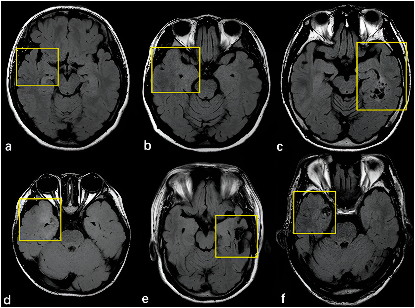
The brain MRI revealed focal cortical dysplasia (FCD) in the left temporal lobe. The image shows multiple MRI slices (a–f), highlighting findings of adjacent cortex in patients with FCD:
- (a) Right hippocampal atrophy without adjacent cortex abnormality.
- (b) Right hippocampal hyperintensity and thickened temporal cortex.
- (c) Ganglioglioma (GG) medial to left temporal lobe with normal adjacent cortex.
- (d) Right temporal pole thickening and indistinct gray–white matter junction.
- (e) Right frontal AVM without visual cortex alteration.
- (f) Cavernous malformation (CCM) in right temporal lobe with cortical hyperintensity.
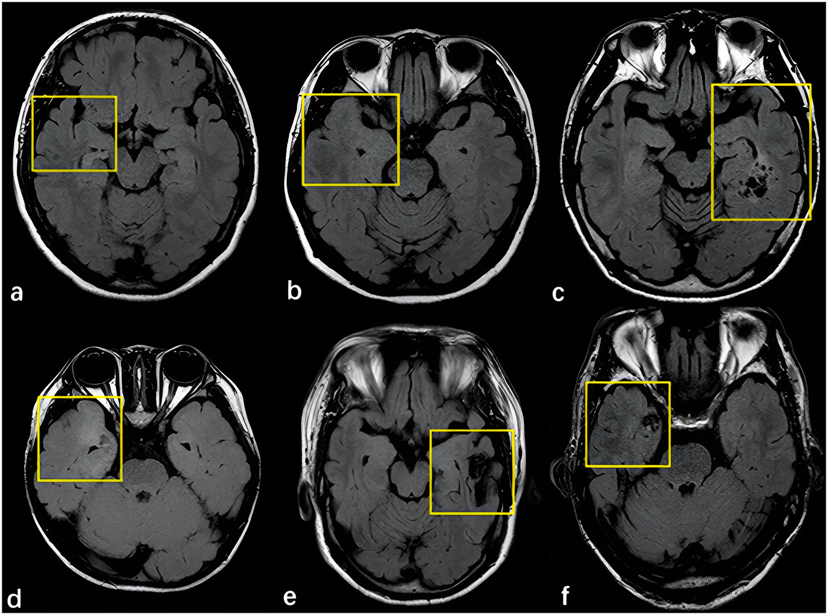
Figure: MRI slices demonstrating various patterns associated with focal cortical dysplasia (FCD) and related cortical abnormalities. Yellow boxes highlight areas of interest across right and left temporal lobes.
Understanding Focal Cortical Dysplasia
FCD is a common cause of epilepsy in children and young adults. It's a developmental brain anomaly where neurons fail to migrate correctly. These abnormal areas can trigger seizures and may not be visible on CT or standard MRI without high-resolution imaging.
Benefits of MRI in Pediatric Neurology
- No radiation exposure — safer for growing brains.
- High-resolution 3D imaging of the cortex and deep brain structures.
- Can detect subtle malformations, tumors, or injuries.
- Can be paired with fMRI and spectroscopy for additional data.
Challenges in Pediatric MRI
- Young children may require sedation to stay still during the scan.
- Specialized pediatric imaging protocols are necessary.
- Interpretation requires neuroradiologists familiar with age-specific anatomy.
Outcome and Follow-Up
• The patient responded well to initial medication.
• Further seizure episodes decreased in frequency and intensity.
• He continues regular follow-ups with neurology and imaging surveillance.
Conclusion: The Role of MRI in Pediatric Seizure Workups
MRI plays a vital role in identifying the underlying cause of seizures in children, especially when other evaluations are inconclusive. Its clarity, safety profile, and diagnostic value make it a critical tool in modern pediatric neurology.