
MRI Uncovers Pituitary Tumor in a Patient with Visual Loss
- Category: Radiology
- Date: 07-08-2025
Introduction: Vision as the First Clue
Visual symptoms such as blurring or field deficits can sometimes signal more than just ophthalmologic issues. The pituitary gland, nestled near the optic chiasm, can exert pressure on visual pathways when tumors grow. MRI is the gold standard for evaluating this region.
Case Presentation
A 42-year-old female presented to an ophthalmologist with complaints of gradually worsening peripheral vision over the past 2 months. She also reported occasional headaches and irregular menstrual cycles. Visual field testing revealed bitemporal hemianopia — loss of vision in the outer halves of both visual fields.
Initial Workup
- Visual acuity: Normal
- Fundoscopy: No papilledema
- Hormonal panel: Elevated prolactin level, low FSH and LH
- MRI brain with sellar protocol was ordered to assess for a pituitary mass
MRI Findings
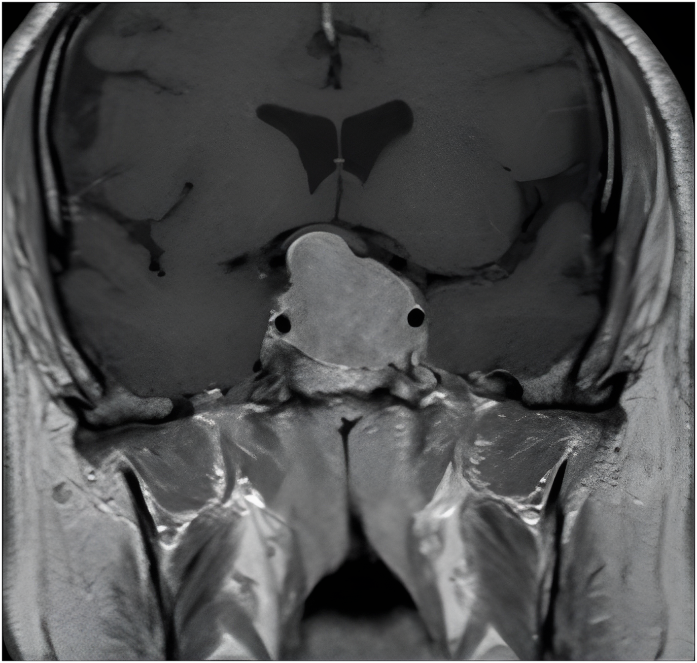
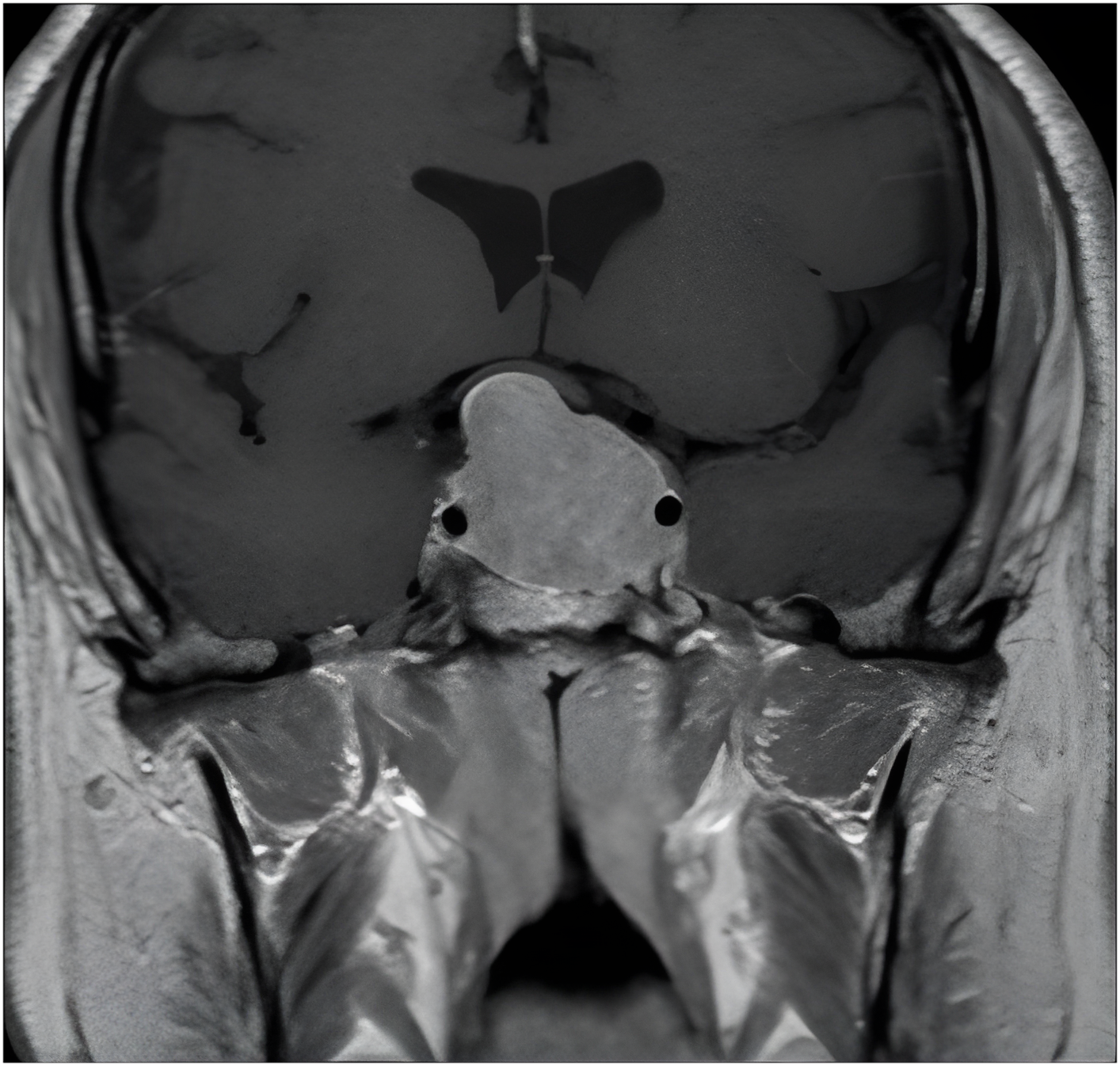
- A **2.5 cm pituitary macroadenoma** was seen occupying the sella turcica and extending superiorly.
- The tumor compressed the optic chiasm, explaining the visual field defect.
- No cavernous sinus invasion was seen.
- The lesion enhanced uniformly with gadolinium.
Diagnosis: Prolactin-Secreting Pituitary Macroadenoma (Prolactinoma)
- Based on the size, hormonal profile, and imaging, the diagnosis of prolactinoma was confirmed.
Management
- Initiated on **dopamine agonist therapy (cabergoline)** to shrink the tumor and normalize prolactin levels.
- Regular follow-up with endocrinology and ophthalmology.
- Repeat MRI planned after 3 months to assess tumor regression.
Why MRI Was Indispensable
- High-resolution imaging of the sellar and parasellar region
- Differentiates between microadenomas (<10 mm) and macroadenomas
- Evaluates optic chiasm, cavernous sinus, and internal carotid proximity
- Dynamic contrast sequences help assess lesion vascularity and enhancement
Patient Outcome
- After 3 months, the patient’s visual fields showed significant improvement.
- MRI confirmed a reduction in tumor size to 1.5 cm.
- Menstrual cycles returned to normal, and prolactin levels normalized.
Conclusion: MRI Guides Targeted Treatment
This case illustrates how MRI can reveal intracranial causes of visual loss that may otherwise be misattributed. Timely diagnosis of a pituitary tumor enabled medical management and visual recovery, avoiding surgical intervention.