
CT Brain Scan Reveals Subdural Hematoma in Elderly Fall Patient
- Category: Radiology
- Date: 05-09-2017
Introduction: The Hidden Danger After a Fall
Falls are a leading cause of injury in older adults. Even minor head trauma in the elderly can lead to serious complications like subdural hematoma (SDH). CT brain imaging is the first-line tool for prompt diagnosis. This case illustrates how early imaging led to life-saving intervention.
Case Presentation
An 82-year-old male was brought to the emergency department by his caregiver after a fall at home the night before. He had tripped over a rug and hit his head, but did not lose consciousness. The next morning, he appeared more confused and sluggish.
Clinical Assessment
- GCS: 14/15
- BP: 140/86 mmHg, HR: 82 bpm
- Neurological exam: Slight right-sided weakness, slurred speech
- No external signs of head injury
- Due to his age and symptoms, a **non-contrast CT brain** was immediately ordered.
CT Findings
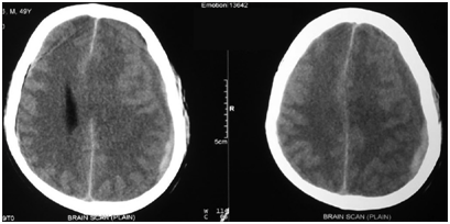
- CT showed a **left-sided crescent-shaped hypodense collection** along the cerebral convexity, consistent with a **subacute subdural hematoma**.
- Mild midline shift (~4 mm) noted, but no herniation.
- No skull fracture or intracerebral bleeding.
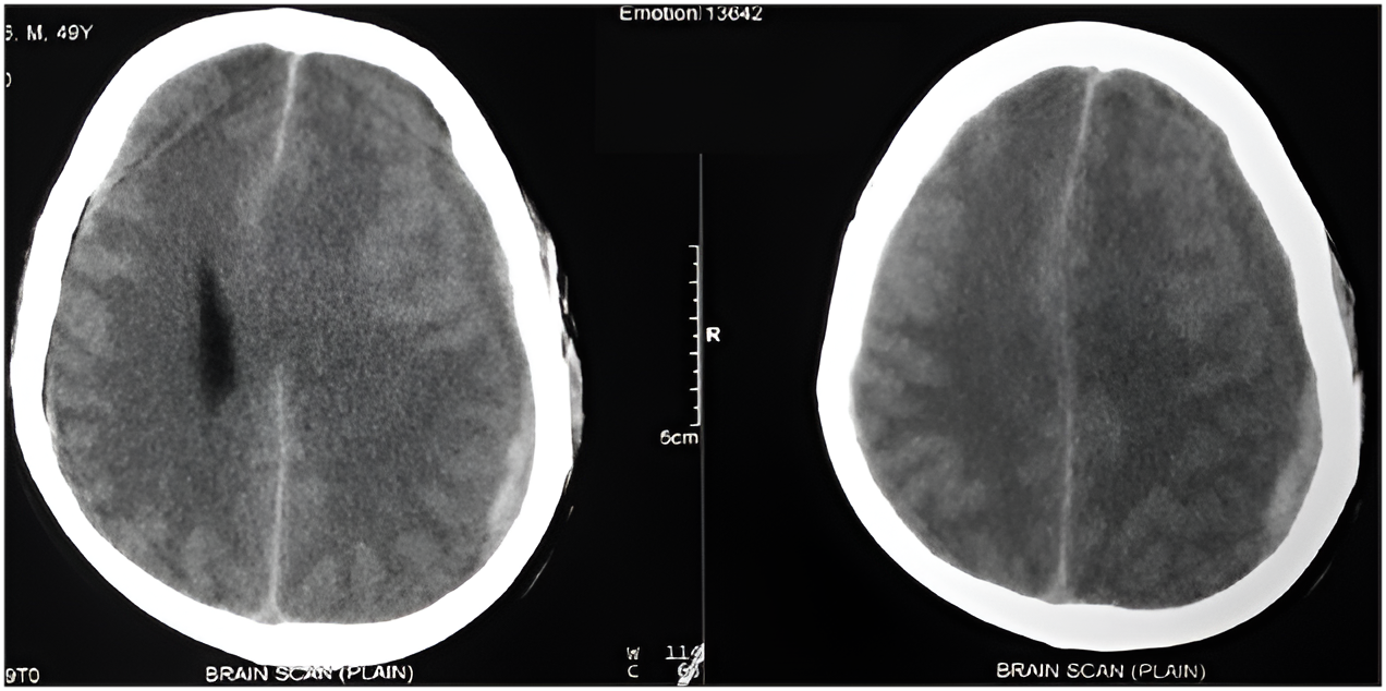
Diagnosis: Subacute Subdural Hematoma
The diagnosis confirmed that the patient's neurological symptoms were due to a developing subdural hematoma from the fall.
Management and Outcome
- Neurosurgery was consulted urgently.
- Given the mild mass effect and stable vitals, conservative management was chosen with close monitoring.
- Patient admitted to neuro ICU.
- Over the next 72 hours, neurological status improved with supportive care.
- Repeat CT after 1 week showed reduction in hematoma volume.
Why CT Was Critical
- Rapid, accessible, and ideal for detecting blood in and around the brain
- Differentiates acute, subacute, and chronic SDH by density characteristics
- Can reveal midline shift, herniation risk, and other associated injuries
Discussion: Subdural Hematoma in the Elderly
- The elderly are more prone due to cerebral atrophy and fragile bridging veins.
- Symptoms may be subtle or delayed — confusion, lethargy, headache, or weakness.
- Even low-impact trauma should trigger neuroimaging in at-risk individuals.
Conclusion: Don’t Dismiss Minor Head Trauma
CT scans are indispensable in evaluating head injuries in older adults. This case demonstrates how prompt imaging and conservative neurosurgical care helped avoid long-term deficits from a seemingly minor fall.