
MRI Diagnoses Multiple Myeloma with Vertebral Involvement
- Category: Radiology
- Date: 07-08-2025
Introduction: When Bone Pain Is a Sign of Malignancy
Multiple myeloma is a plasma cell malignancy that frequently affects the axial skeleton. Patients often present with back pain due to vertebral involvement or pathologic fractures. MRI is the most sensitive modality to detect early marrow changes and spinal cord compression.
Case Presentation
A 67-year-old male presented with persistent mid-back pain and fatigue for two months. He denied trauma or heavy lifting. He also complained of unintentional weight loss and frequent infections.
Initial Workup
- CBC: Anemia (Hb 9.8 g/dL)
- ESR: 105 mm/hr, Calcium: Elevated at 11.3 mg/dL
- Serum protein electrophoresis: M-protein spike
- X-rays showed lytic lesions in thoracic spine and ribs
- MRI spine was ordered to assess extent of disease
MRI Findings (Updated for Image)
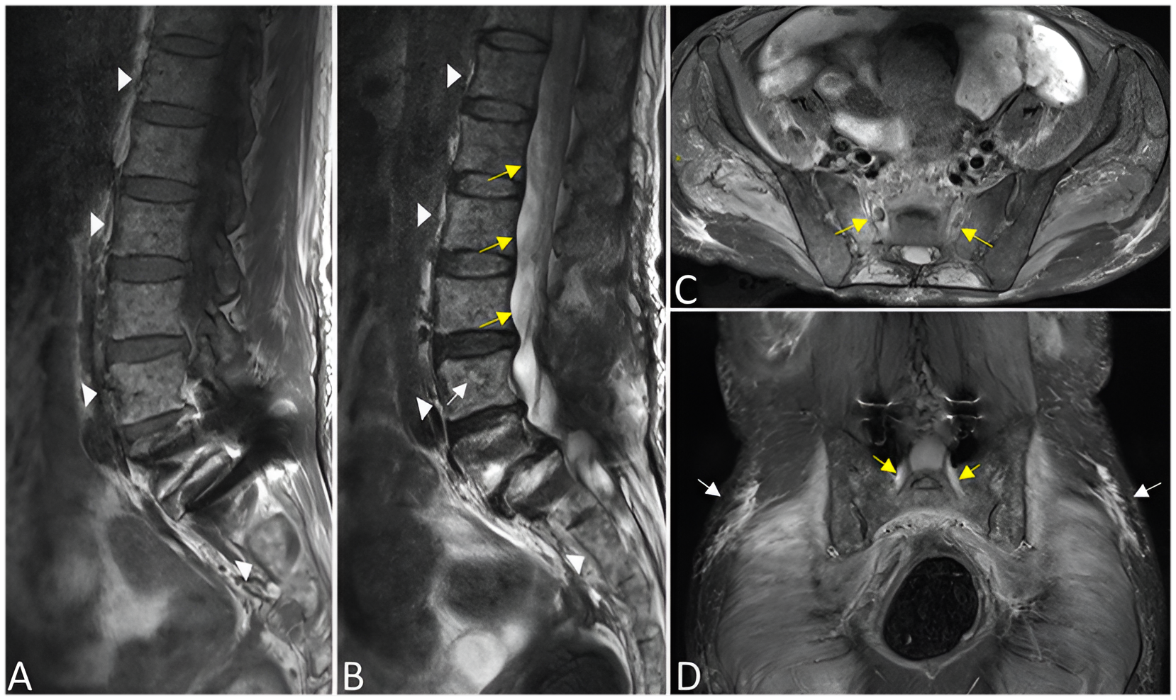
• Sagittal T1-weighted images (Figure A) showed multiple hypointense marrow-replacing lesions throughout the lumbar vertebral bodies.
• Sagittal T2-weighted images (Figure B) revealed corresponding hyperintense lesions with vertebral body height loss at L2–L4, suggesting compression changes.
• An epidural soft tissue component was noted, with spinal canal narrowing at L3–L4.
• Axial T2 and coronal STIR images (Figures C and D) revealed bilateral sacroiliac involvement and soft tissue extension, consistent with extensive marrow infiltration.
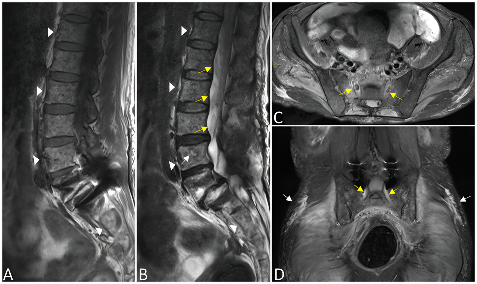
Figure: Multiplanar MRI of the lumbar spine and pelvis in multiple myeloma.
(A) Sagittal T1-weighted showing hypointense vertebral lesions (white arrowheads).
(B) Sagittal T2-weighted with hyperintense lesions and mild compression fractures (yellow arrows).
(C) Axial T2 showing bilateral sacral marrow lesions (yellow arrows).
(D) Coronal STIR showing bilateral sacroiliac involvement and epidural soft tissue (yellow and white arrows).
Diagnosis: Multiple Myeloma with Spinal Involvement
MRI confirmed widespread lytic disease and a vertebral fracture secondary to multiple myeloma.
Management
- Started on chemotherapy (bortezomib + dexamethasone + lenalidomide)
- Bisphosphonates for bone protection
- Neurosurgical evaluation for spinal stability; opted for conservative management with bracing
- Hematology follow-up for stem cell transplant eligibility
Why MRI Was Critical
- Superior sensitivity to detect bone marrow infiltration
- Identifies soft tissue extension, spinal canal compromise, and fracture risk
- Essential for staging and treatment planning in multiple myeloma
Patient Outcome
• Responded well to initial chemotherapy
• Back pain significantly improved with bracing and radiation
• Repeat MRI after 3 months showed regression of soft tissue lesions
Conclusion: MRI Unveils the Silent Bone Invasion
MRI is indispensable in the diagnosis and management of multiple myeloma involving the spine. It allows early detection, accurate staging, and prevention of catastrophic spinal complications in patients with minimal symptoms.