
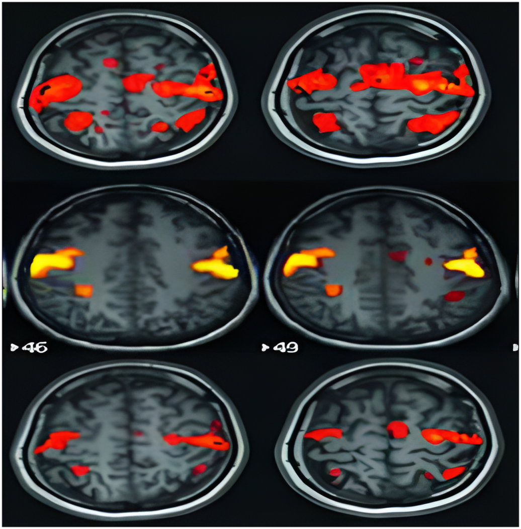
Functional MRI (fMRI): Seeing the Brain in Action
- Category: Radiology
- Date: 05-09-2017
Introduction: Imaging the Living Brain
While traditional MRI gives detailed pictures of brain anatomy, functional MRI (fMRI) goes one step further — it captures brain activity in real time. fMRI has transformed neuroscience and neurology by offering a non-invasive method to visualize which parts of the brain are working, when, and how.
What is Functional MRI?
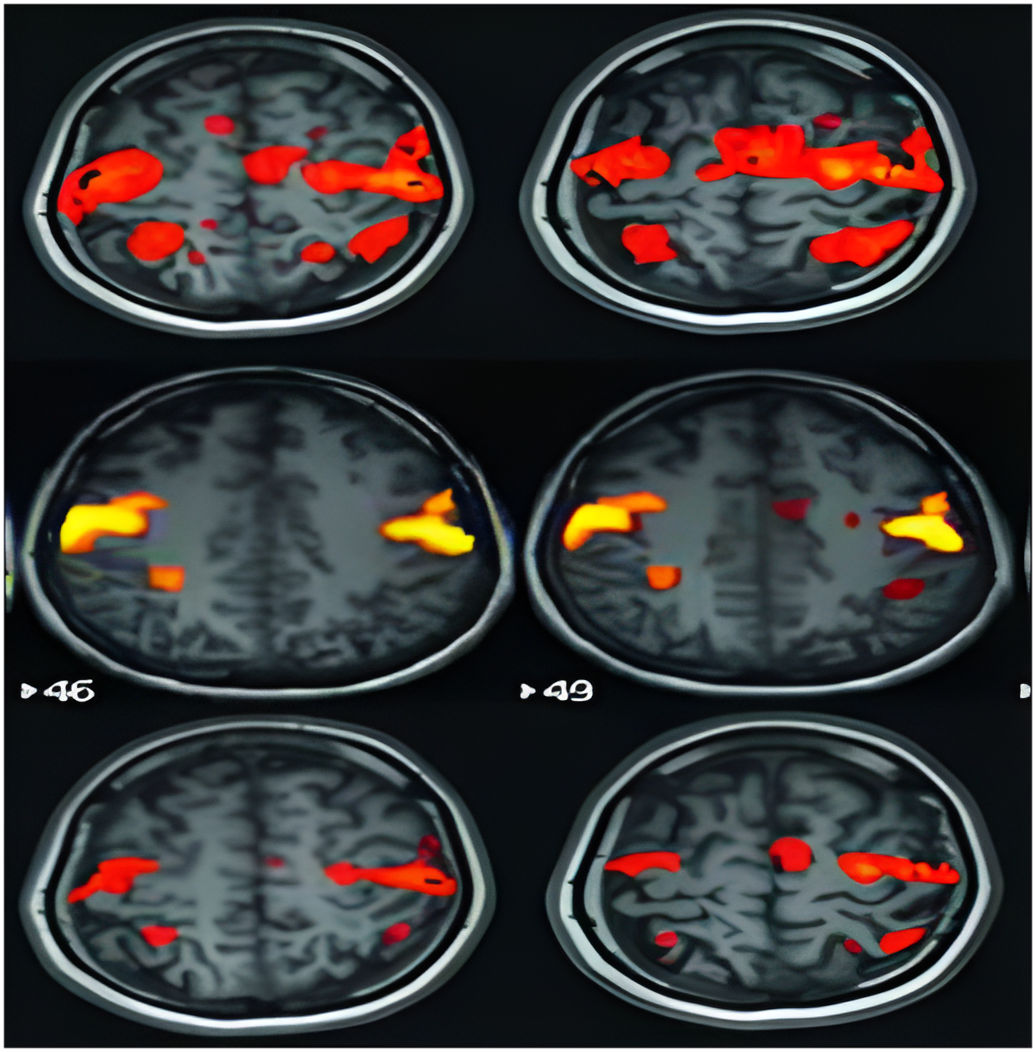
Functional MRI, or fMRI, measures changes in blood flow within the brain. It is based on the principle that active brain areas require more oxygen. As neurons fire, nearby blood vessels increase blood supply — a process called the BOLD (Blood Oxygen Level Dependent) response. fMRI detects these subtle changes and maps them onto brain images.
How fMRI Works
- A standard MRI machine is used, with specialized software.
- The patient is asked to perform simple tasks (e.g., tapping fingers, looking at images, solving math problems).
- The machine detects changes in oxygenated vs deoxygenated blood in specific brain regions.
- These changes are color-coded and overlaid on anatomical brain images.
Clinical Applications of fMRI
- **Neurosurgery Planning**: Identify and preserve essential brain regions before removing tumors.
- **Stroke Rehabilitation**: Understand which areas are compensating post-stroke.
- **Epilepsy Evaluation**: Pinpoint seizure-generating zones.
- **Cognitive Disorders**: Study memory and decision-making in Alzheimer’s or Parkinson’s disease.
- **Psychiatry Research**: Explore emotional regulation and mental health disorders.
Benefits of fMRI
- **Non-invasive** and does not use radiation.
- **Real-time insight** into brain function.
- **Improves surgical outcomes** by preserving language or motor areas.
- **Guides personalized treatment plans** for neurological conditions.
Limitations of fMRI
- **Not a diagnostic tool** for specific diseases on its own.
- **Motion sensitivity** — even slight head movement can distort results.
- **Requires cooperation** from patients to perform tasks.
- **Expensive** and not available at all imaging centers.
Research and Emerging Applications
- **Brain-Computer Interfaces (BCIs)**: fMRI is being used to develop systems that let paralyzed patients control devices using thoughts.
- **Lie Detection and Legal Use**: Still controversial, but under investigation.
- **Mental Health Insights**: fMRI studies are exploring the biological basis of depression, anxiety, and PTSD.
Case Example
A patient with a low-grade brain tumor near the motor cortex is scheduled for surgery. Pre-operative fMRI is performed while the patient moves their fingers and toes. The imaging identifies motor areas to avoid during tumor removal. As a result, the tumor is successfully resected without impairing movement.
What to Expect During an fMRI
- The procedure is similar to a regular MRI.
- You’ll lie in the scanner while following prompts on a screen.
- You may be asked to solve puzzles, view images, or respond using a hand-held button.
- No injections or contrast agents are usually required.
Conclusion: Mapping the Mind in Motion
Functional MRI is a powerful tool for understanding how the brain works — not just what it looks like. Whether guiding surgery, studying cognition, or advancing neuroscience, fMRI opens a window into the dynamic human mind like never before.