
Brain Tumor Case: The Role of MRI in Early Detection
- Category: Radiology
- Date: 05-09-2017
Introduction: Headaches That Won’t Go Away
Headaches are one of the most common symptoms in outpatient care. But sometimes, they can point to something more serious. This case study highlights how MRI played a crucial role in detecting a brain tumor at an early stage — when treatment options were most effective.
Case Presentation
A 36-year-old woman presented to her neurologist with persistent headaches for three months. They were dull, mostly on the left side, and occasionally accompanied by blurry vision. Neurological examination was normal. No prior history of migraines or trauma.
Initial Evaluation and Clinical Suspicion
Given the patient’s age and new, progressive headache pattern, neuroimaging was warranted. Although a CT scan can be a useful first-line test, her physician opted for MRI due to its higher sensitivity for soft tissue abnormalities and posterior fossa imaging.
MRI Findings
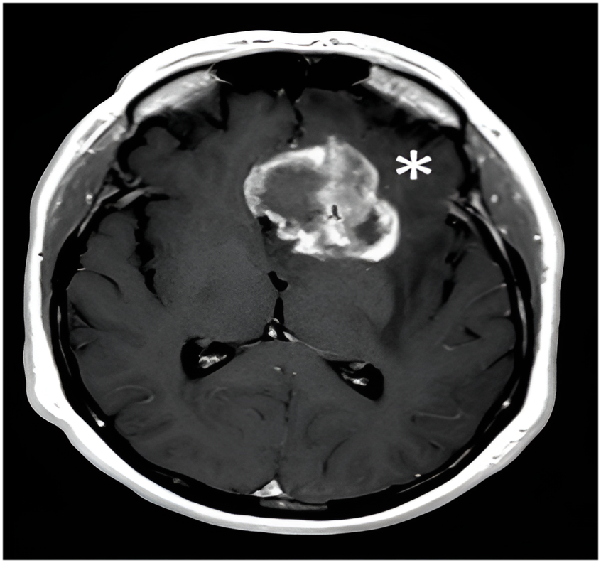
- An MRI of the brain was performed with and without gadolinium contrast.
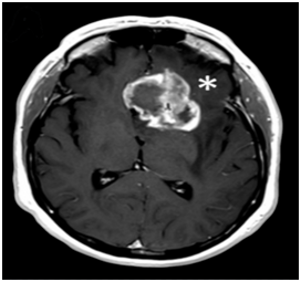
- Imaging revealed a mass in the left frontal lobe.
- The lesion was well-circumscribed, enhancing with central necrosis and surrounding vasogenic edema, and caused mild midline shift.
- Features suggested a Glioblastoma multiforme (GBM).
Additional Workup
- MR Spectroscopy: Showed mildly elevated choline-to-creatine ratio, supporting a neoplastic process.
- Functional MRI: Used to map language centers due to the tumor’s proximity to Broca’s area.
- Neurosurgical evaluation was requested for biopsy and resection planning.
Diagnosis: GBM
Surgical Outcome and Follow-Up
- The tumor was successfully resected with minimal residual.
- Post-operative MRI confirmed gross total resection.
- The patient experienced no neurological deficits.
- She is under close MRI surveillance every 6 months.
- Adjuvant therapy was deferred pending signs of progression.
Why MRI Was Crucial
- **CT scan might have missed the lesion** or mischaracterized it.
- **MRI provided detailed views** of the tumor’s location, effect on nearby structures, and internal characteristics.
- Functional MRI allowed safe surgical planning, avoiding speech impairment.
Lessons from the Case
- Don’t ignore persistent, unexplained headaches — especially when new in onset after age 30.
- MRI is the gold standard for detecting brain tumors, particularly in non-emergency, subtle cases.
- Early detection allows better outcomes and broader treatment options.
Conclusion: MRI as a Lifesaving Diagnostic Tool
This case underscores the power of MRI in diagnosing brain tumors at a treatable stage. By offering unparalleled clarity and functional insights, MRI enables early intervention — often before life-altering symptoms occur.